

Editorial: Naël El Kastani
In our health-related professions, understanding the human body is based on the analysis of its anatomical structures. Yet, this internal richness—this complex network of organs, tissues, muscles, blood vessels, and nerves—remains difficult for our patients to perceive.
So, why not integrate clear, beautiful, and relevant human anatomy diagrams into your professional space? What if an illustrated anatomical model became a clinical, relational, and even emotional tool? 🧠
An anatomical diagram, available on our site En Chair et En Os, is more than just a visual aid: it is a powerful means to better locate, understand, and act upon parts of the body.
Revealing the invisible: the pedagogical function of the anatomical diagram
Representing the body's unconscious: a key for patients
Many patients arrive for consultations with a vague idea of their internal anatomical function. The location of an organ, the connection between a muscular system and a nerve, or the position of a blood vessel are often unknown to them. Yet, for a patient to commit to a course of treatment, they must understand its foundations.
📌 The Occipito-Thyro-Thymo-Pericardial Fascia Chart is a reference image to illustrate this. This vertical structure connects key areas of the body: the cranial cavity, thyroid gland, anterior mediastinum, and pericardium.
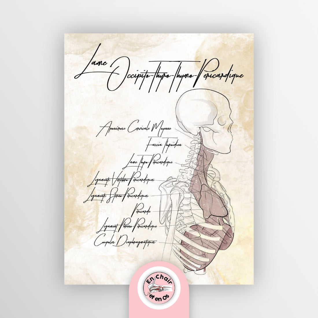
➡ We are talking here about a central anatomical axis, at the junction between the central nervous system, the respiratory system, and the cardiovascular system.
💬 Explaining to a patient that their palpitations, neck pain, or shortness of breath can stem from tissue tension along this fascia changes everything.
🧩 This anatomical model helps to compose a global vision, connect the dots, and promote the integrative approach you practice in your office.
Training and transmitting: a tool in the service of practitioners
In multidisciplinary offices or during clinical internships, it is fundamental to pass on knowledge of anatomy and physiology. A good anatomy diagram then becomes a structuring element of continuing education.
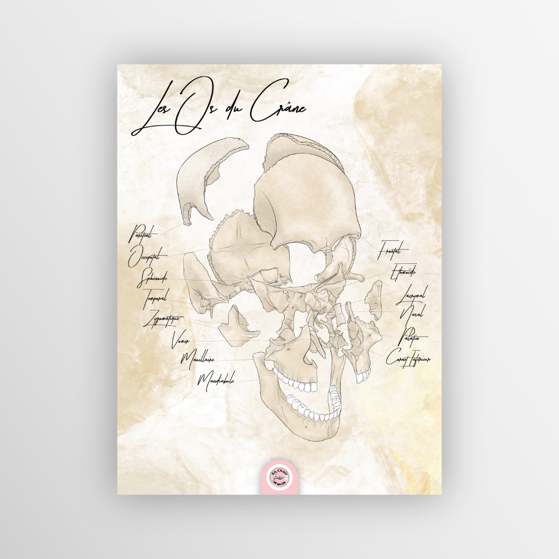
👁️ Let's take the Skull Bones Chart:
-
It offers a precise view of the cranial skeleton, with its osseous sutures, neurovascular passage zones, and connections with the cervical spine.
-
It allows one to locate muscular anchor points, areas of cerebral irrigation, or the cerebral functions associated with each zone.
🎓 This anatomical chart becomes a pedagogical tool for understanding how dysfunction in a skull bone can have neurological or even visceral repercussions.
✅ It is no longer just a simple "image," but a teaching product that makes anatomical information accessible, visual, and structured.
Creating a body-space dialogue: when the image structures the environment
Organizing space around the trunk: coherence and logic of the center
Your medical office or treatment room says something about your therapeutic approach. If you think of the human body as a coherent whole, why not reflect that in the visual organization of your room?
👉 The Thoracic Diaphragm Chart is perfect for anchoring your space in the idea of centrality.

-
This respiratory tissue, located between the thorax and the abdomen, constitutes a major anatomical crossroads.
-
It forms the link between the lungs, heart, stomach, ribs, spine, and the vessels of the trunk.
💨 Integrating it into your consultation space serves as a reminder that the center of the body is also a functional and emotional center. The diaphragm plays a crucial role in managing stress, postural tone, and even visceral motility.
📍 The image creates a centering point, a stable reference around which your therapeutic presence is built.
Giving the neck a role: posture, emotion, and nervous network
The neck is one of the most highly innervated and taxed areas of the human body. It acts as a junction node between the head, trunk, and upper limbs. It is also a mirror of our emotional state.
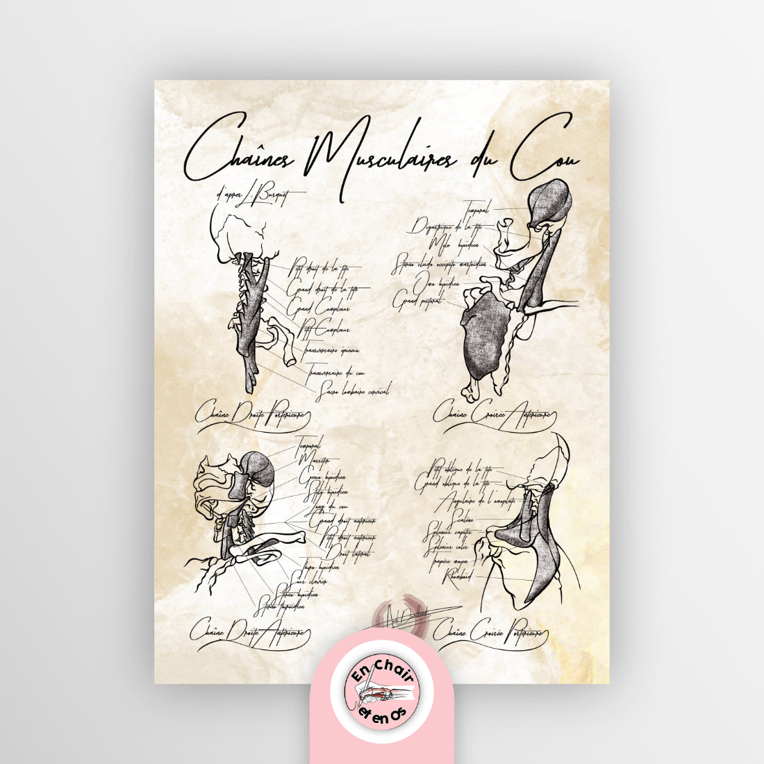
The Neck Muscle Chains Chart is a powerful anatomical reference for explaining to a patient that their headaches, dizziness, or tingling in the hands can originate from cervical tension.
-
One can see the large anterior muscles, the posterior elements, as well as the structuring of the neck into functional chains.
-
This includes the scalene muscles, the upper trapezius, and the sternocleidomastoid—all essential in managing posture, mobility, and the brain's neurovascular network.
💬 A diagram here is not just a support. It is a communication tool. It makes the invisible obvious; it justifies manual treatment, breathing exercises, or postural reprogramming.
Giving meaning to movement: charts to support action
The psoas: pivot of movement and internal balance
The iliopsoas muscle is probably one of the most important, yet most underestimated, muscles in the human muscular system. It connects the lumbar spine to the femur, passes through the abdomen, and inserts deeply into the pelvis.
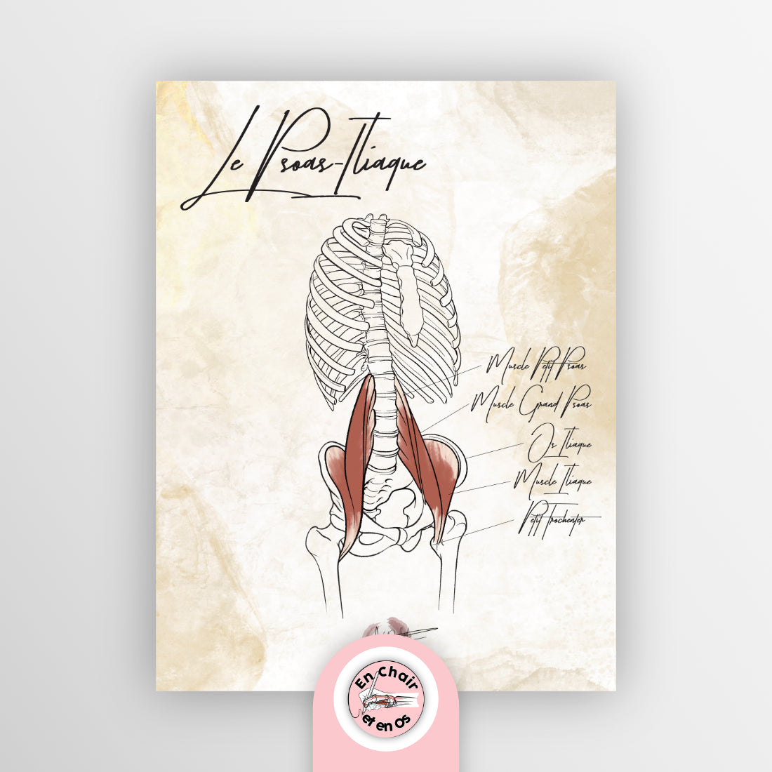
➡ The Anatomical Chart - the Iliopsoas reveals its full functional complexity.
-
It is involved in hip mobility, but also in respiration, posture, and lower back pain.
-
It influences the digestive system due to its proximity to abdominal organs, and participates in the autonomic nervous system.
💡 Showing this anatomical content during a session allows the patient to realize why their back, belly, and even their emotions are linked by one and the same muscle.
📌 This repositions the role of anatomy in daily clinical practice and transforms a local treatment into a global care approach.
The body in action: embodying anatomy through movement
Let's conclude with a chart that embodies body dynamics, the Cyclist Chart. It portrays anatomy in action, and this is of paramount importance.
🚴♀️ This chart highlights the muscle chains engaged, the joint angles, the mechanical limits… and above all, it connects the static anatomical model to the dynamic movement of the body.
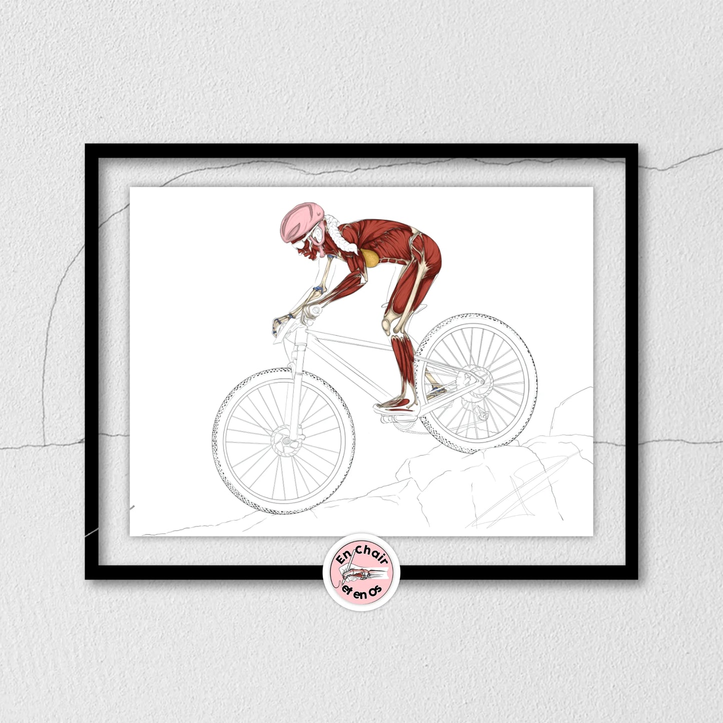
It is ideal for a practitioner who:
-
works in athletic reconditioning or lower limb rehabilitation (knee, hip, ankle),
-
offers functional sports tracking,
-
or addresses postural prevention.
🎯 This chart shows that the body is not a sum of parts, but a coordinated, mobile, living, and adaptive system.
✅ Conclusion: a tool, a mission, a transformation
The anatomical diagram is more than just an image: it is a clinical tool, a vector for transmission, a pedagogical foundation, an element of visual structure, and sometimes even a trigger for therapeutic change.
In every page, every diagram, and every chart on our site En Chair et En Os, lies a way to better grasp, locate, and act upon the human body.
📣 What about you? Which diagram will you integrate into your practice to transform the way you treat your patients?