

Written by: Naël El Kastani
Anatomy is our common language, the one that connects all healthcare professionals. But in daily practice, how do we convey this language of the human body to the patient? How do we make it visible, understandable, and alive? An anatomical drawing displayed in a consultation room, or used actively by the practitioner, is much more than a visual tool: it is a medium for dialogue, a means of learning, and a gateway to understanding how the human body functions.
Drawing as a mediator in the caregiver-patient relationship
Translating medical language with a visual aid
Human anatomy, when explained only with words, can leave the patient lost. But with a drawing of the human body? That is a different story. Thanks to an anatomical chart, a line, an angle, or a muscle becomes legible. The pose of an anatomical figure or an illustrated model allows the patient to understand, learn anatomy, and read their own body through it.
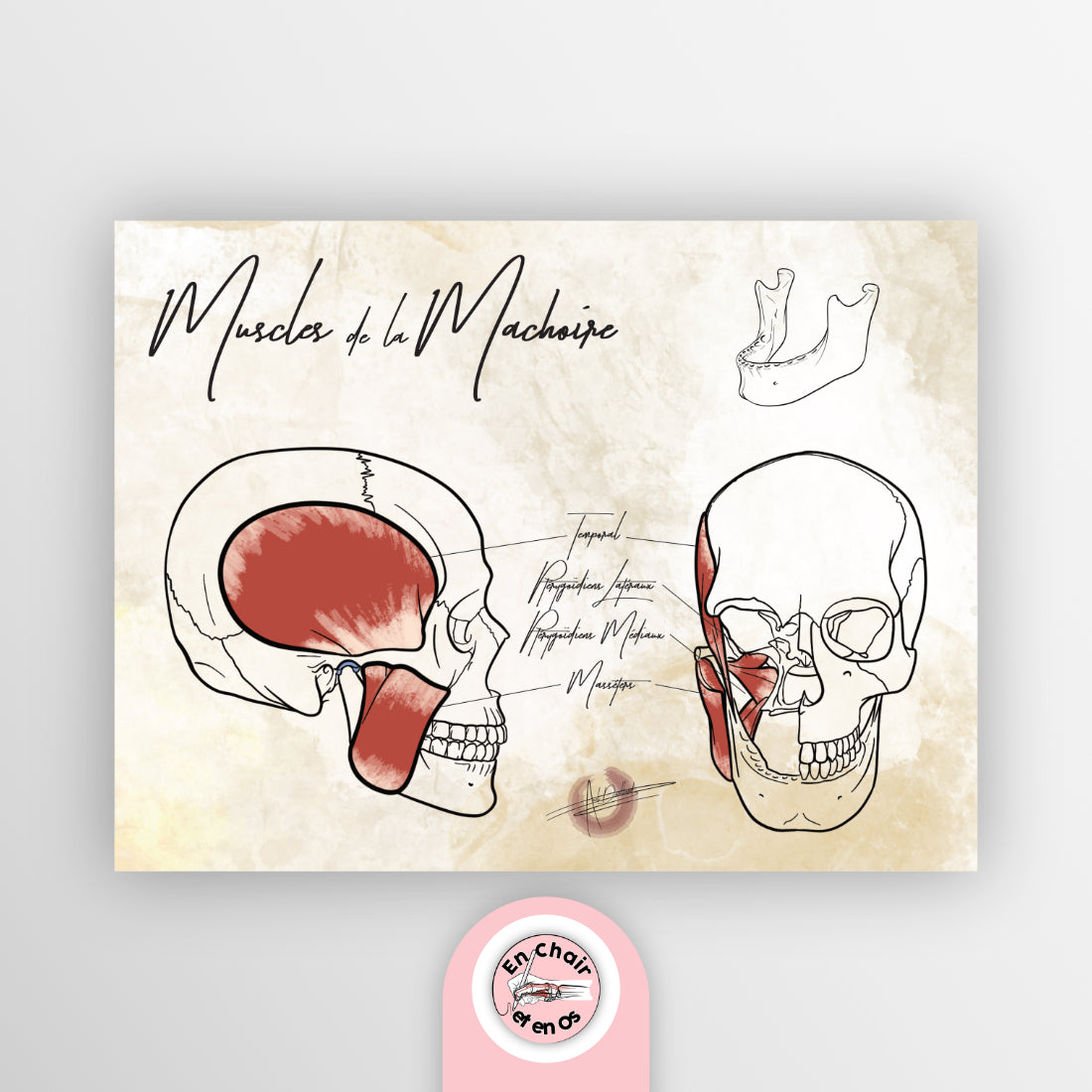
👉 Example: the Anatomical Chart - Jaw Muscles, ideal for explaining TMJ disorders, this chart makes the complexity of the head, face, and deep muscles visible. It offers a simple and effective way to explain muscle functions, imbalances, and pain. It also serves as a foundation for suggesting drawing methods or clinical exercises, halfway between anatomy for sculptors, artists, and therapists.
✏️ This image becomes an educational tool, a method for learning anatomy that is accessible to both the patient and the practitioner.
Humanizing the consultation through imagery
When a practitioner draws, or shows a drawing, they step out of technical discourse to create a human exchange. It is a simple way to offer a moment of shared understanding, an experience where the patient learns by looking, asking questions, and taking an interest in their own body.

👉 Example: the Anatomical Chart - Football - Zidane, both aesthetic and precise, grabs attention. It allows for discussions about movement, lower limb joints, posture types, and at-risk zones. In the context of rehabilitation, it helps to show the concrete effects of a sprain, a joint blockage, or a muscle imbalance.
💬 Here, the image becomes a starting point for learning, prevention, and even therapeutic follow-up.
A clinical tool for movement and pathology analysis
Understanding postural imbalances at a glance
In the treatment of chronic pain, musculoskeletal disorders, or postural pathologies, a well-placed classic anatomy drawing makes it possible to compare, explain, and visualize the differences between the anatomical model and the patient’s clinical reality.
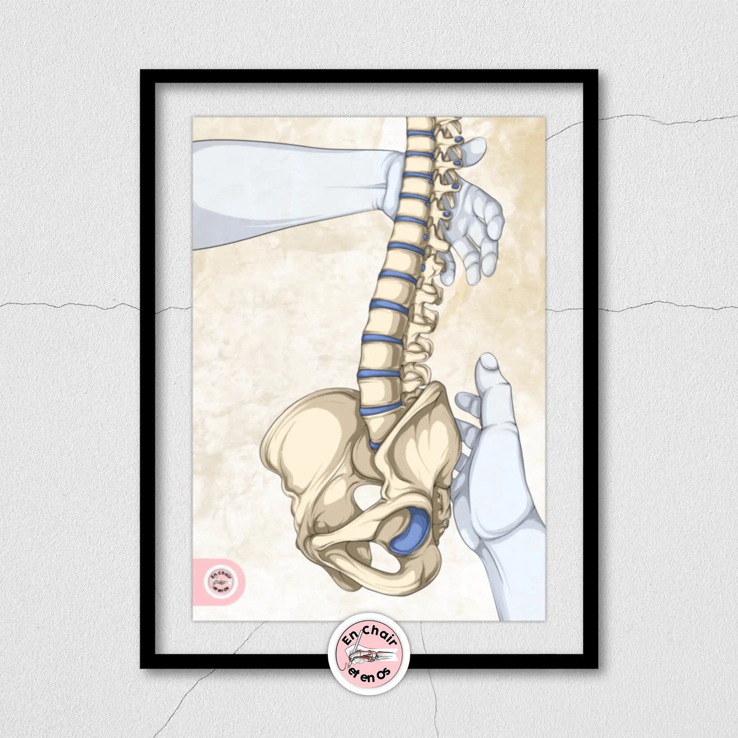
👉 Example: the Chart - Spine and Pelvis – Pelvic Girdle, essential for any postural consultation, details the relationship between the pelvis and the spine. It is perfect for drawing the body, explaining natural proportions and the clinical deviations that need correction.
📏 A chart like this helps guide the patient toward a better understanding of their body, in direct connection with the practice of drawing, trunk anatomy, and even strengthening exercises.
Targeting therapeutic techniques
Before a manual treatment, an injection, or mobilization, showing the area, drawing the structure, and offering a clear visualization helps reassure the patient and structure the intervention. The patient knows where we are working, why, and how.

👉 Example: the Chart - The Rider, very comprehensive, highlights the effects of prolonged sitting, particularly for people who work in a seated position or practice horse riding. It shows compression zones, back muscles, and the posterior chain in their anatomical function.
🪑 An excellent visual diagnostic tool, and a work guide for suggesting postural correction methods.
Drawing as a training and cultural tool for the practitioner
Learning anatomy beyond words
A book offers pages to read, but a drawing also engages the hand. For a healthcare professional, drawing anatomy—even quickly—allows one to rediscover structure, fix details in the mind, and memorize the relationships between body parts. It is a full-fledged anatomy learning exercise.
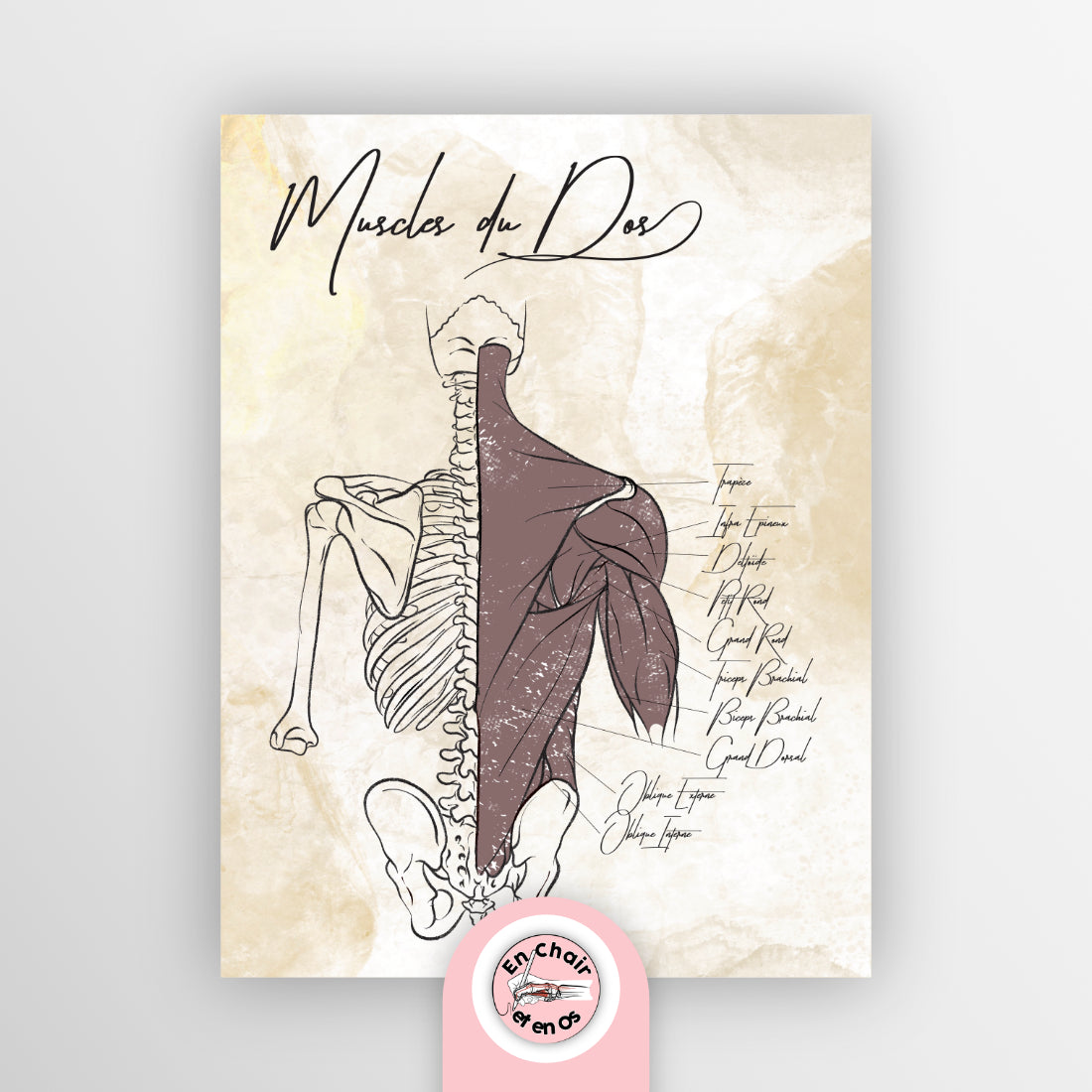
👉 Example: the Chart - Back Muscles, a precision tool for any healthcare professional or artist. It shows the different muscular layers, attachments, movements, and key proportions for drawing figures or offering treatment based on anatomical observation.
✍️ A study method that combines drawing practice, anatomy books, artistic anatomy, and clinical tools. Perfect for learning how to draw the human body, or how to treat it.
Creating a cultural space in the healthcare setting
A well-chosen anatomical chart can also be an inspiration. It becomes an invitation to discover the body differently, to share a passion for anatomy, and to provide educational content to the patient—why not a paperback book, a video, an online illustration, or a training sketch?
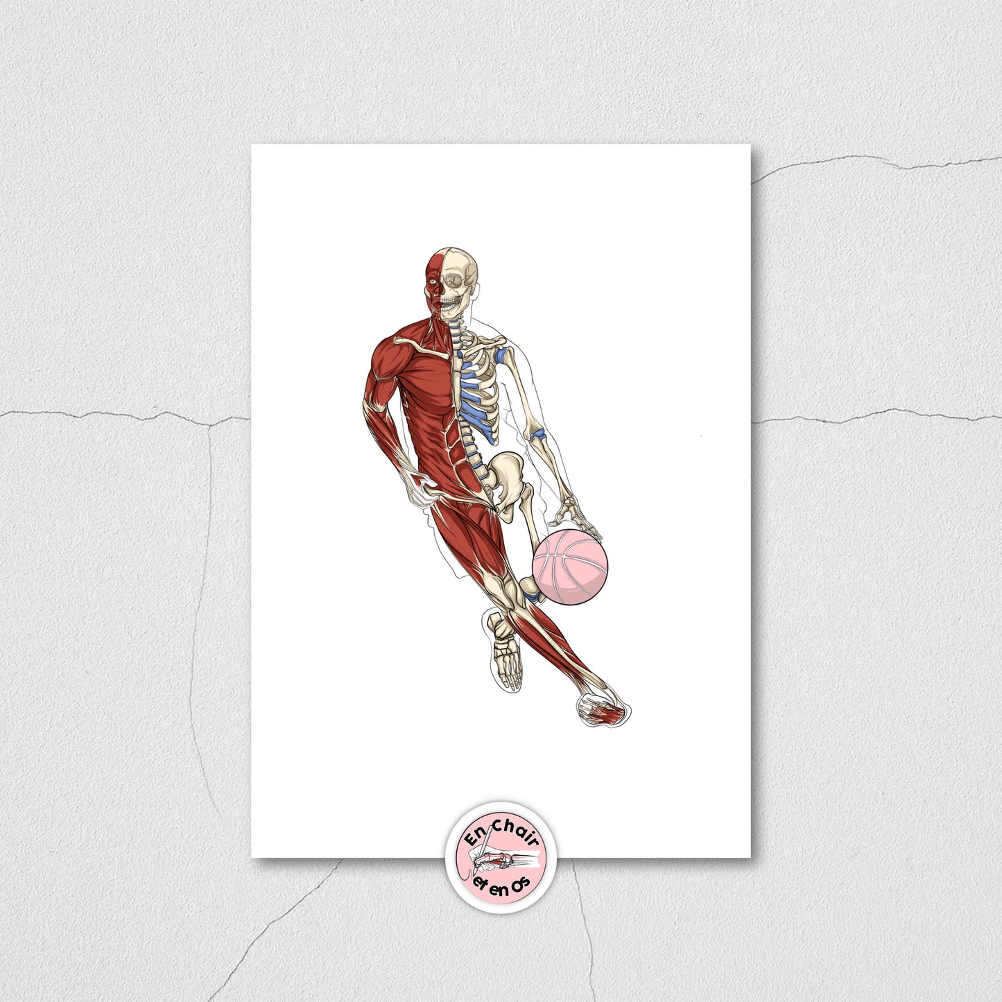
👉 Example: the Chart - Basketball, colorful and dynamic, perfectly shows upper body movement, rotation gestures, and the upper limbs in action. It is an ideal chart for discussing joint angles, upper limb anatomy, and the impact of sporting movements.
🏀 It stimulates curiosity, offers a visual aid for explaining pain or injuries, and bridges the gap between sport, art, and medicine.
Conclusion
The anatomy of the human body is too precious to be left in a closed book. By drawing, showing, and explaining with images, we invite the patient to understand, learn, and experience their body as a legible system.
The collection of charts available on our site En Chair et en Os offers high-quality products: paperback formats, online illustrations, detailed comments, and anatomical models, all designed to help both the caregiver and the learner.
🧩 In short, drawing anatomy means reading the body in depth, treating through pedagogy, and sharing living knowledge.