Skip to product information
1 of 2

En Chair et en Os

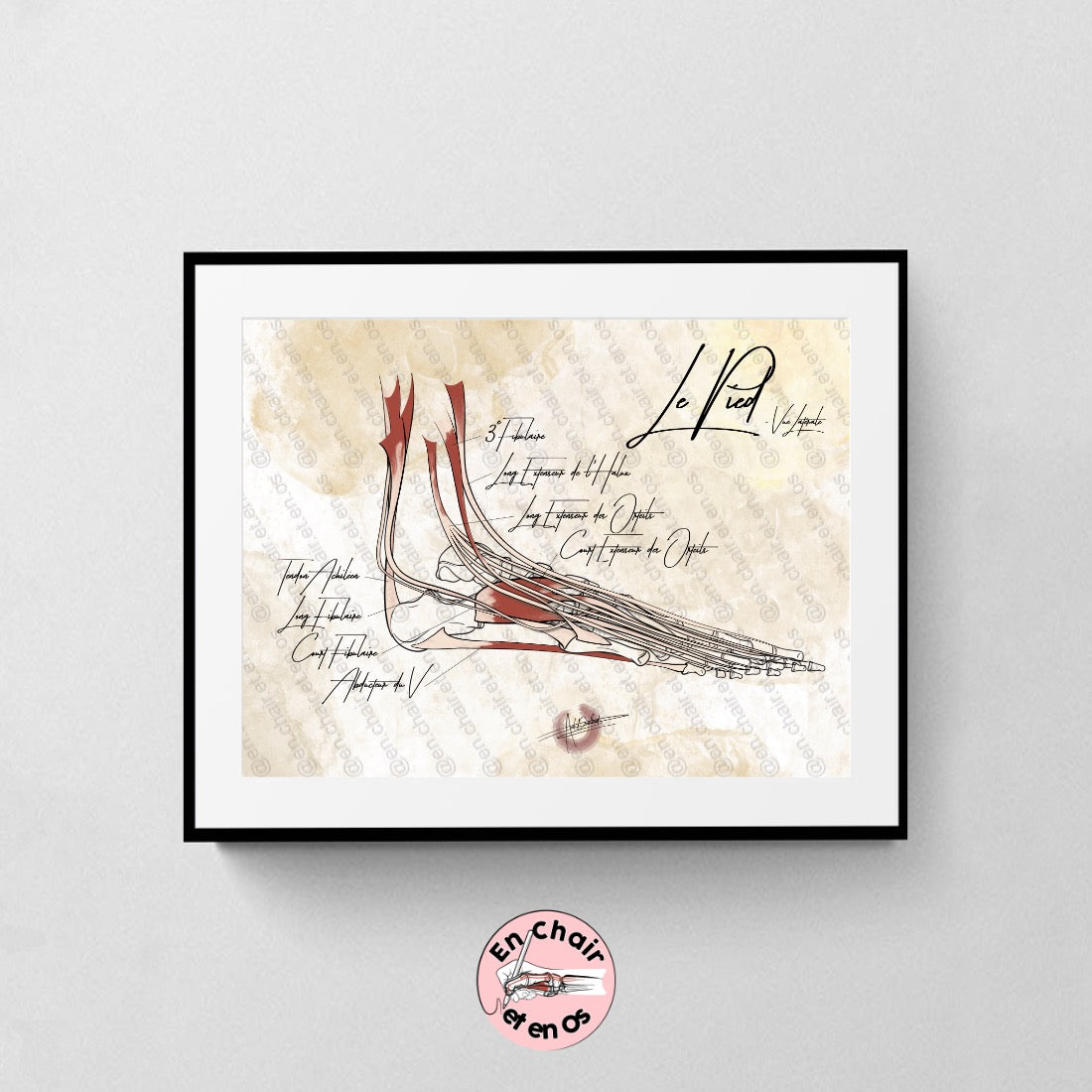
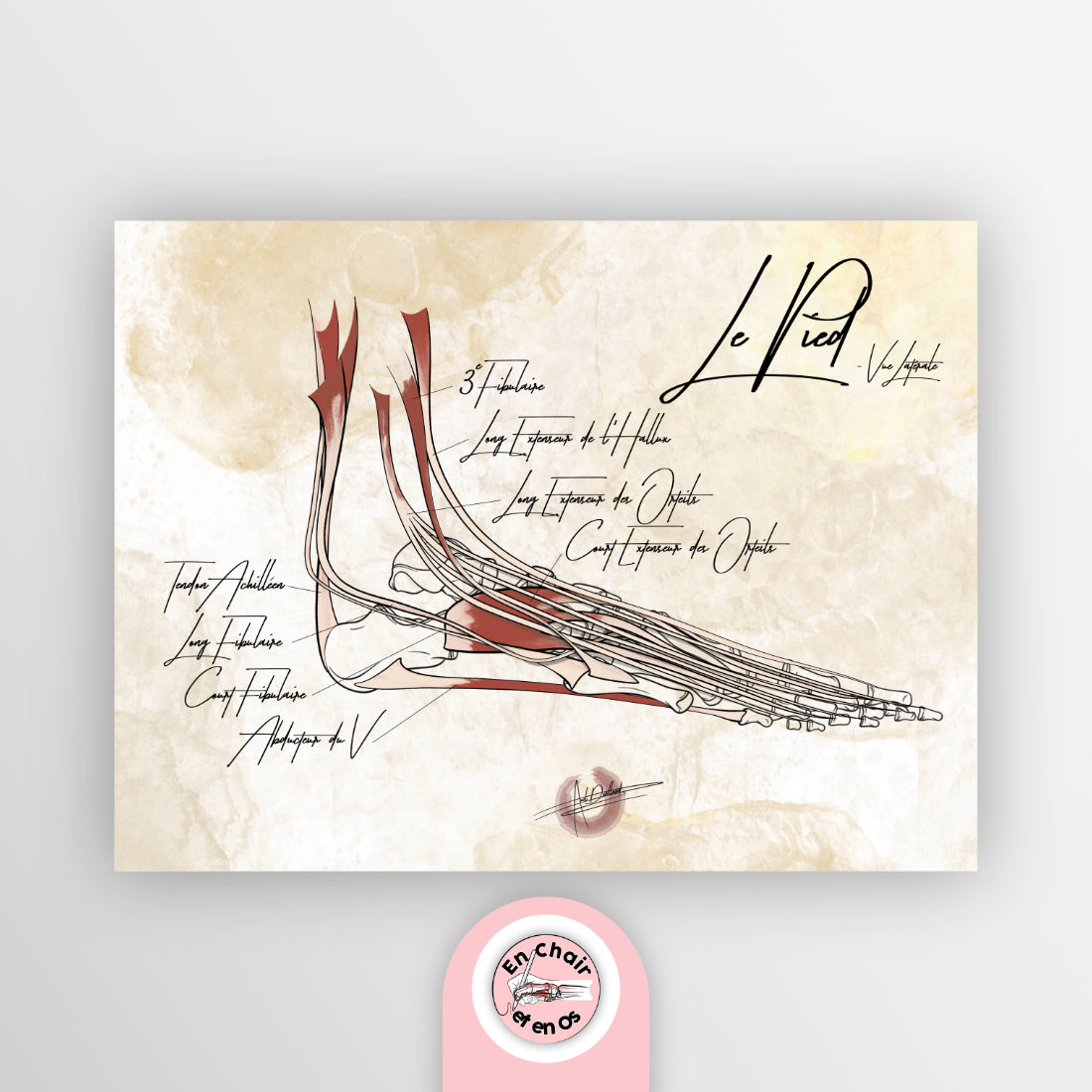
Anatomical chart of the foot – Lateral view

Anatomical chart of the foot – Lateral view

Regular price €45,00 EUR
Regular price Sale price €45,00 EUR
Sale Sold out
Tax included. Shipping calculated at checkout.

Format

Hand-illustrated by Axel Devillard, creator of En Chair et en Os.

Anatomical chart of the foot, lateral view: the bones and muscles of the foot

The foot bears the entire weight of the body with every step. This anatomical chart of the foot shows it in profile, from the external side, with its bones and muscles hand-drawn. A clear view to understand how the foot articulates with the ankle and how it stabilizes during walking.

What the chart shows

  • The bones of the foot seen in profile: tarsus, metatarsus, and phalanges, up to the ankle
  • The muscles and tendons of the lateral edge of the foot and the ankle

An ideal visual aid for explaining an ankle sprain, tendinopathy, or pain on the outer edge of the foot.

Who is this chart for?

  • Podiatrists: to decorate the office and support the explanation of the podiatric assessment
  • Physiotherapists and osteopaths: to illustrate ankle and foot rehabilitation
  • Sports doctors and foot surgeons
  • Students of podiatry, physiotherapy, osteopathy, medicine, and sports science, to review foot anatomy

Specifications

  • Hand-illustrated by Axel Devillard, creator of En Chair et en Os
  • Two sizes: 30 × 40 cm (A3 equivalent) and 40 × 60 cm (A2 equivalent)
  • Printed in France on 300 gsm recycled paper
  • Frame not included
  • Order prepared by hand and shipped within 3 to 5 business days

Complete the decor of your podiatry clinic

Frequently asked questions

How many bones are in the foot?

The foot has 26 bones: 7 tarsal bones (including the talus and calcaneus), 5 metatarsals, and 14 phalanges. In addition, there are small sesamoid bones under the head of the first metatarsal.

Why a lateral view of the foot?

The lateral view shows the profile of the arch, the ankle, and the external edge of the foot, where most sprains occur. It is the most intuitive view for a patient.

Which size should I choose?

The 30 × 40 cm is suitable for a desk or treatment room. The 40 × 60 cm is readable from a distance and becomes the centerpiece of a clinic wall.

Choose your size and receive your anatomical chart of the foot.

View full details

Customer Reviews

Overall rating: 5.0 / 5 from 2 reviews.

Excellente communication avec Axel. D’une gentillesse extraordinaire et efficace. Les visuels sont juste magnifique.

Planche anatomique du Pied - Vue latérale -

Store owner replied: Merci Tanguy, je suis plus que touché par votre retour très motivants !

Excellent poster d’anatomie, à la fois précis et esthétique. Les détails anatomiques sont bien représentés et les légendes très lisibles. Un support pédagogique de grande qualité au cabinet !

Planche anatomique du Pied - Vue latérale -